
Human anatomy lab manuals provide a comprehensive guide for understanding the structure and function of the body through hands-on dissection exercises, including cat dissections, offering practical experience.
Overview of Human Anatomy Lab Manuals
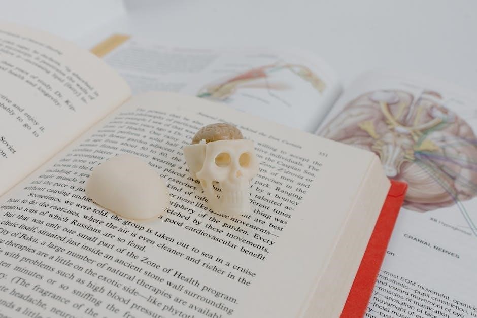
Human anatomy lab manuals are comprehensive guides designed to enhance understanding of the body’s structure and function. They typically include detailed dissection exercises, such as cat dissections, to provide hands-on learning experiences. These manuals cover all major body systems, offering step-by-step instructions, full-color illustrations, and clinical application questions. They serve as essential tools for students, blending practical and theoretical knowledge to foster deeper anatomical comprehension and preparation for real-world scenarios.
The Role of Cat Dissections in Anatomy Education
Cat dissections play a crucial role in anatomy education by providing students with a hands-on learning experience. Cats have anatomical structures similar to humans, making them ideal for studying muscles, organs, and body systems. These dissections offer a practical understanding of human anatomy, enhancing textbook knowledge and preparing students for clinical applications while fostering appreciation and engagement in the learning process through direct observation and exploration.
Preparation for Cat Dissections
Preparing for cat dissections involves gathering materials, following safety protocols, and understanding ethical guidelines to ensure a respectful and educational experience for anatomy students.
Materials and Tools Needed
The essential tools for cat dissections include scalpels, forceps, dissecting scissors, and gloves. Preserved cat specimens, lab coats, and goggles are required for safety. Additional materials like trays, magnifying lenses, and anatomy charts enhance the learning experience, ensuring students can explore and identify structures effectively while maintaining a sterile and organized workspace.
Safety Protocols and Precautions
Wear gloves, goggles, and lab coats to protect against biohazards. Handle sharp tools with care, ensuring proper ventilation in the lab. Dispose of waste in designated containers and avoid eating or drinking nearby. Follow proper handwashing procedures and adhere to lab rules to maintain a safe environment for all participants during cat dissections and related activities.
Ethical Considerations and Lab Etiquette
Adhere to ethical standards when handling specimens, ensuring respectful treatment of cat dissection materials. Maintain a clean workspace, label all tools, and avoid unnecessary damage to tissues. Collaborate courteously with lab partners, minimize disruptions, and follow instructions to promote a professional and respectful learning environment. This fosters mutual respect and accountability among students and instructors alike.

Muscular System Dissection
The muscular system dissection focuses on identifying and understanding cat muscles, emphasizing their structure, function, and similarity to human anatomy, providing hands-on experience in anatomical study.
Skeletal Muscles of the Cat
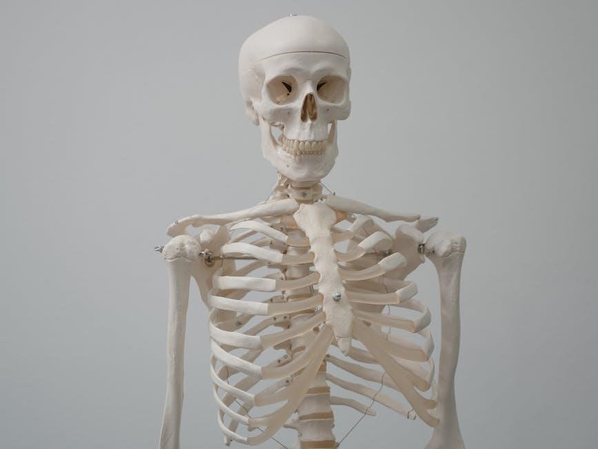
Skeletal muscles in cats are well-developed, making them ideal for studying muscle structure and function. These muscles, attached to bones, enable movement and locomotion. Their anatomy closely resembles human skeletal muscles, providing a comparative learning tool. Dissection allows detailed examination of muscle groups, such as flexors and extensors, highlighting their roles in movement and overall feline physiology.
Abdominal Muscles and Their Functions
The abdominal muscles of a cat are layered, consisting of the external and internal oblique, transversus abdominis, and rectus abdominis. These muscles support the spine, protect internal organs, and facilitate movements like walking or running. Their structure and function closely mirror human abdominal muscles, making them an excellent model for comparative anatomy study in educational dissections.
Muscles of the Lower Limbs
The lower limb muscles of a cat include the quadriceps, hamstrings, and gastrocnemius, which are essential for movement and balance. These muscles enable flexion and extension of the knees and ankles, facilitating locomotion. Dissection of these muscles provides insight into their structure, function, and role in movement, offering a valuable comparison to human anatomy and aiding in the understanding of muscle mechanics and their interconnections.
Ventral Body Cavities
Ventral body cavities in cats, including thoracic and abdominal regions, house vital organs like the heart, lungs, and digestive structures, mirroring human anatomy for educational dissection purposes.
Thoracic Cavity and Its Contents
The thoracic cavity in cats contains the heart and lungs, protected by the rib cage. During dissection, the diaphragm separating thoracic and abdominal cavities is removed to expose these organs, providing insights into their structure and function, closely resembling human anatomy for educational purposes.
Abdominal Cavity and Organ Identification
The abdominal cavity in cats houses essential organs like the liver, stomach, intestines, pancreas, spleen, and kidneys, protected by the peritoneum. Dissection allows detailed examination of their structure and relationships, mirroring human anatomy. Students learn to identify and understand the spatial arrangement of these organs, enhancing their comprehension of digestive, urinary, and endocrine systems through hands-on exploration.
Pelvic Cavity and Reproductive Structures
The pelvic cavity in cats contains reproductive organs, including the uterus, ovaries, and vagina in females, and the bladder, prostate, and testes in males. These structures are smaller but anatomically similar to humans. Dissection allows students to explore the spatial relationships between these organs, providing insights into the urinary and reproductive systems while highlighting the interconnectedness of pelvic anatomy in mammals.
Nervous and Circulatory Systems
The nervous and circulatory systems are essential for maintaining life, with the nervous system controlling functions and the circulatory system transporting oxygen and nutrients throughout the body.
Structure of the Nervous System
The nervous system consists of the central nervous system (CNS), including the brain and spinal cord, and the peripheral nervous system (PNS), comprising nerves and ganglia. In cat dissections, students examine the spinal cord, nerve roots, and peripheral nerves, gaining insights into the structural organization of the nervous system and its role in controlling bodily functions. This hands-on approach enhances understanding of neural pathways and their significance in human anatomy.

Key Components of the Circulatory System
The circulatory system includes the heart, arteries, veins, and capillaries, working together to transport blood throughout the body. In cat dissections, students observe the heart’s chambers, blood vessels, and valves, gaining insights into the system’s structure and function. This hands-on approach helps understand how the circulatory system maintains tissue health and overall bodily functions, mirroring human anatomy for educational purposes.
Digestive and Respiratory Systems
The digestive and respiratory systems are essential for nutrient absorption and oxygen exchange. Cat dissections provide a hands-on understanding of these interconnected systems, mirroring human anatomy for educational purposes.
Digestive System Anatomy
The digestive system, including the stomach, small intestine, liver, and pancreas, is meticulously examined during cat dissections. These structures closely resemble human anatomy, allowing students to study digestive processes and organ functions in detail. The dissection process enhances understanding of nutrient absorption and waste elimination, providing a hands-on learning experience that aligns with human physiological mechanisms.
Respiratory System Components
The respiratory system components include the trachea, bronchi, lungs, and diaphragm, which are meticulously studied during cat dissections. These structures closely mirror human anatomy, enabling students to explore the pathway of air through the trachea into bronchi and alveoli for gas exchange. Dissection provides a detailed understanding of how these organs function in breathing and oxygenation, offering practical insights into respiratory processes and their essential roles in overall physiology, making it a valuable learning experience.

Clinical Applications and Case Studies
Cat dissections in anatomy labs bridge the gap between theoretical knowledge and real-world clinical applications, enabling students to explore human health and disease through hands-on learning and case analysis.
Applying Anatomy Knowledge to Clinical Scenarios
Cat dissections provide practical experience in understanding human anatomy, enabling students to apply their knowledge to real-world clinical scenarios. By studying feline anatomy, learners can better grasp human anatomical structures and their functions. This hands-on experience enhances their ability to diagnose and treat conditions, preparing them for careers in medicine and healthcare. The similarities between cat and human anatomy make dissections a valuable tool for clinical training and decision-making.
Case Studies in Anatomy Education
Case studies in anatomy education bridge theory and practice, offering real-world insights. Using cat dissections, students analyze anatomical structures and apply knowledge to clinical scenarios. These exercises enhance understanding of muscle functions, organ systems, and their interconnections. Practical experience with feline anatomy provides a foundation for interpreting human anatomical variations, fostering critical thinking and observational skills essential for future medical professionals.
Lab Safety and Biohazard Protocols
Lab safety and biohazard protocols are crucial for minimizing risks during dissections. Essential measures include wearing gloves, goggles, and ensuring proper specimen handling and disposal. Emergency preparedness is vital.
Proper Handling of Specimens
Proper handling of cat specimens is essential for safety and respect. Wear gloves, goggles, and lab coats to prevent exposure. Use appropriate tools for dissection, ensuring precise and careful techniques. Maintain a clean, organized workspace to avoid contamination. Store specimens in sealed containers when not in use. Follow ethical guidelines and institutional protocols for specimen care and disposal.
Disposal of Biohazardous Materials
Dispose of biohazardous materials, such as cat specimens and tissues, in designated red biohazard bags. Seal bags securely and label them as biohazardous waste. Autoclave or incinerate materials according to lab protocols. Store waste in a designated area until pickup. Ensure all disposal methods comply with institutional and environmental safety regulations to minimize health risks and environmental impact.
Emergency Procedures in the Lab
In case of emergencies, such as chemical spills or injuries, immediately alert lab personnel. Use eyewash stations or safety showers if exposed to hazardous materials. For cuts, apply pressure and seek medical attention. Evacuate the area if there’s a fire or gas leak and activate alarms. Keep emergency contact numbers and first aid kits accessible to ensure prompt response and minimize risks during dissections or lab activities.
Human anatomy lab manuals with cat dissections provide invaluable hands-on experience, enhancing understanding of bodily structures. Future editions could integrate virtual dissection tools and 3D models to complement traditional methods, offering a hybrid learning approach. Expanding clinical application questions and incorporating real-world case studies will further prepare students for professional scenarios, ensuring a comprehensive and modern anatomical education.